Decoding Posture Series: Post 2
How the Lower Leg Shapes Whole‑Body Alignment
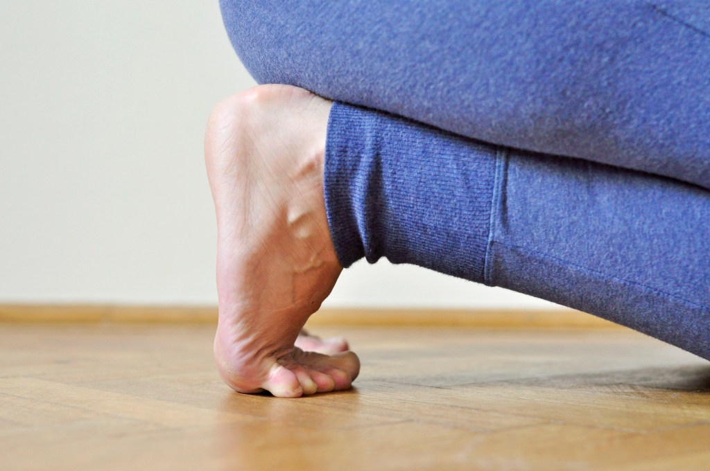
When we look at posture, the ankles and knees often get overlooked — yet they’re two of the most revealing joints in the entire kinetic chain. They tell us how the body manages load, absorbs force, and adapts to the patterns that begin at the feet. The first time I ever thought about reduced ROM in the ankle and witnessed the impact on the rest of the body was when I started teaching yoga. Some clients struggled to sit back into Child’s Pose; inability to go all the way down into Malasana (deep squat to the floor); knee drift and foot turnout in Utkatasana (Chair Pose/squat).
Why ankles and knees matter in posture assessment
These joints act as the body’s natural shock absorbers. When they move well, the whole system feels smoother and more efficient. When they stiffen, collapse, or rotate excessively, the body compensates above.
Common patterns you’ll see:
- knees drifting inward (valgus)
- knees bowing outward (varus)
- ankles collapsing medially
- ankles rolling outward
- limited dorsiflexion affecting gait and squat patterns
These are common adaptations.
How foot mechanics influence the knees
A pronated foot often creates an inward spiral up the leg. A supinated foot often creates an outward spiral. But both influence knee tracking.
What to observe:
- Does the knee track over the second toe?
- Does one knee behave differently from the other?
- Does the knee collapse inward during load?
- Does the client “lock” their knees in standing?
Neutral as a behaviour
Neutral knee alignment isn’t just a fixed position: it’s the ability to move through neutral with ease.
Look for:
- smooth transitions
- balanced weight distribution
- the ability to bend without collapsing
- the ability to straighten without discomfort
Simple awareness tests
- Stand on one leg — what happens at the knee?
- Try a slow mini‑squat — does the knee drift?
- Walk barefoot — do the knees rotate inward or outward?
Biomechanics: what’s happening under the surface
A useful way to think about the ankle–knee relationship is rotation. When the foot pronates to accept load, the arch lowers and the tibia often follows with a small amount of internal rotation. If the hip can’t control that rotation (or the ankle/foot collapses too quickly), the knee may drift inward. With a more supinated foot, the tibia tends to rotate outward and the knee may sit more “bowed” (varus) or struggle to absorb force smoothly.
Dorsiflexion matters because it’s one of the main ways we decelerate bodyweight during walking, stairs, and squat patterns. If the ankle can’t flex, the body comensates: the foot may turn out, the arch may roll inwards, the heel may lift early, or the knee may travel medially to find range. Those compensations are useful clues in assessment.
Screening Tell Tale Signs
Squat to stand or a wall squat: early heel lift, foot turnout or valgus. What it might suggest: limited dorsiflexion range or dorsiflexion not well controlled under load.
Shoulder Bridge: the supporting foot “claws” at the mat or rolls in/out, the pelvis drops/hitches, the knee loses stability and alignment as weight shifts during foot switch. What it may suggest: reduced foot tripod organisation and/or a hip control demand for which the client is not yet ready.
Mini case study: knee drift in a squat pattern
Client presents with tendency towards valgus during sit-to-stand test and there is evidence of instability on single-leg tasks. In natural standing posture, you notice that one foot and and the knee on the same side roll inwards slightly. In a slow mini-squat, the tibia appears to internally rotate as load increases, the heel wants to lift early, and the knee tracks the big toe rather than the second toe. A quick knee-to-wall check suggests reduced dorsiflexion compared with the other side. You then have to continue to observe to decide whether it is a foot/ankle ROM issue, foot control issue or hip control issue.
The Knee to Wall Test assesses ankle dorsiflexion by measuring how far the foot can be from a wall while the knee touches it without the heel lifting. Adequate dorsiflexion is essential for proper gait, squatting, and weight-bearing activities. Limited mobility can contribute to conditions like knee pain, overpronation, or plantar fasciitis.
You can help this client by restoring dorsiflexion, improving awareness of the foot tripod and work on hip external rotation/abduction. When you retest the mini-squat or single leg work, hopefully you will see better knee tracking.
Useful Cues
- “Track the knee over the second toe” or “keep the heel heavy as you bend”.
Note: discomfort can have many contributors: if pain is sharp, worsening, or persistent, seek assessment from a qualified health professional.
If you want to deepen your ability to read these patterns in clients, my Postural Assessment course explores posture in much more detail.
